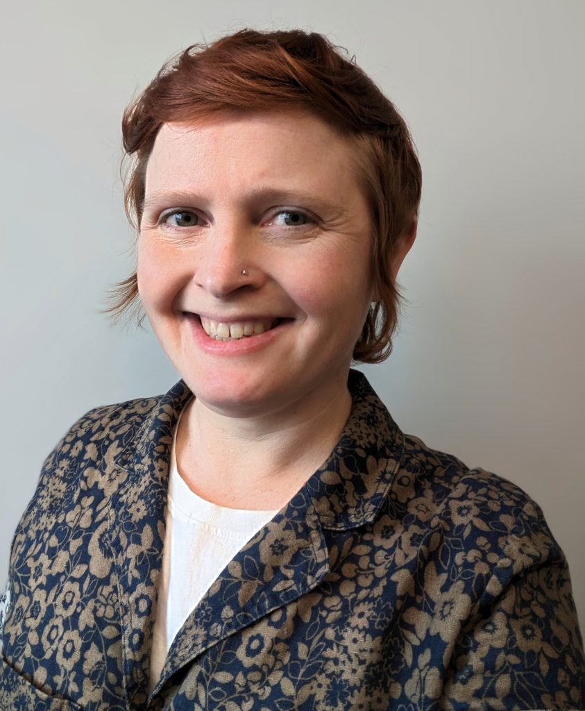
New Faculty Spotlight: Sarah Shelby

Assistant Professor Sarah Shelby has long been fascinated by biology.
“Even back in my years as an undergraduate chemistry major, I was a little bit in awe of the complexity found in biological systems, and have been deeply curious about how complex structures in the cell are fundamentally shaped by basic chemical and physical forces,” she said.
Shelby did her PhD at Cornell University, where she took advantage of a new form of fluorescence microscopy called “super-resolution microscopy.” This method can resolve structures in intact cells as small as 10–20 nanometers, which is ten times smaller than what is possible with conventional fluorescence microscopy. The ability to “see” features so small provided access to the inner workings of complex cellular structures like the plasma membrane, and a new avenue to understand how cells sense and respond to cues from their environment.
“I first used this method in my PhD work to image clustering and dynamics of the IgE receptor, which is the immune receptor in mast cells responsible for allergic responses,” said Shelby.
As a postdoc at the University of Michigan she focused on how interactions between lipids organize plasma membrane components, and how membrane organization impacts signal transduction from cell surface receptors. Lipid-lipid interactions are fascinating because even though they tend to be relatively weak and non-specific compared to interactions between proteins, collectively they lead to non-uniform mixing of the “2D fluid” of the plasma membrane, including its constituent proteins. This effectively compartmentalizes signaling biochemistry on the membrane surface in a similar way that organelles compartmentalize different cellular functions.
“Using super-resolution microscopy, I was able to directly image lipid domains around B cell receptors and show that these domains concentrate key receptor signaling partners in a way that promotes signal transduction,” she said.
She is extending this research at UT.
“I am studying a new receptor system, known as Chimeric Antigen Receptors or CARs,” said Shelby. “CARs are part of a new class of immunotherapy treatments where immune receptors are engineered to direct the immune system to fight cancer. I’m very interested to see if we can use what we know about how plasma membrane structure and organization regulates native immune receptors to investigate how CARs trigger immune responses, and even come up with strategies for how CARs can be improved. We will use super-resolution microscopy to visualize CAR signaling interactions in the plasma membranes of live cells during the initiation of the cellular response.”
Since arriving at UT, she has been building a super-resolution microscope. This is an entirely custom instrument and will be put together as a team effort between Shelby and students in the lab.
“The microscope will be our main experimental tool and I’m excited about involving students in the process because this kind of hands-on experience will give them an opportunity to learn how it works from the bottom up,” she said. “This way, we’ll be in a great position to execute on new ideas and applications for super-resolution microscopy, and push the limits of what the technique is capable of.”
Research in the Shelby lab is interdisciplinary and students will use methods based in a variety of fields including cell biology, biochemistry, biophysics, optics and instrumentation, and quantitative image analysis.
Shelby joined BCMB in February 2023 and is team-teaching BCMB 401 (Biochemistry 1) this fall. Outside the lab and classroom, she is an avid hiker and camper and is looking forward to exploring the Smokies. She also enjoys cooking, gardening, and biking around town.